Click on
 to download a PDF document.
Search within the articles (PDF documents) using Google Search:
Citations
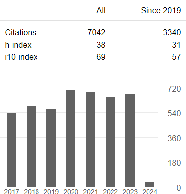
Click on the graph for more details (Google Scholar)
Book chapters
| 4. |
|
Garcia D, Villemain O.
Les promesses de l'échocardiographie ultrarapide. Le corps en images. Les nouvelles imageries pour la santé. CNRS Éditions, Paris, 2022. |
| 3. |
|
Garcia D.
Au cœur du tourbillon cardiaque. Voir l'invisible. (Gex JP, association ARMIR, ed.) Éditions du Puits Fleuri. 2019. |
| 2. |
|
Garcia D, Lantelme P, Saloux É.
Introduction to speckle tracking in cardiac ultrasound imaging. Handbook of speckle filtering and tracking in cardiovascular ultrasound imaging and video. (Loizou CP, Pattichis CS, d'Hooge J, ed.) Institution of Engineering and Technology. 2018. |
| 1. |
|
Garcia D, Durand LG.
Modeling of aortic stenosis and systemic hypertension. Wiley encyclopedia of biomedical engineering. (Metin Akay, ed.) Hoboken: John Wiley & Sons, Inc. 2006. |
Conference papers (selected)
| 6. |
|
Choupani S, Varray F, Gilles B, Béra JC, Garcia D.
Arterial pressure loss from vascular vector flow mapping with conventional color Doppler. IEEE International Symposium on Biomedical Imaging. IEEE. 2023. |
| 5. |
|
Ecarlat P, Perrot V, Carcreff E, Nicolas B, Liebgott H, Garcia D.
Alias-free color Doppler with chirps. IEEE International Ultrasonics Symposium. IEEE. 2022. |
| 4. |
|
Garcia D.
Make the most of MUST, an open-source MATLAB UltraSound Toolbox. IEEE International Ultrasonics Symposium. IEEE. 2021. |
| 3. |
|
Perrot V, Garcia D.
Back to basics in ultrasound velocimetry: tracking speckles by using a standard PIV algorithm. IEEE International Ultrasonics Symposium. IEEE. 2018. |
| 2. |
|
Badescu E, Ambrogio S, Fenner J, Liebgott H, Friboulet D, Garcia D. Vortex ring phantom for investigation of ultrasound vector flow imaging. IEEE International Ultrasonics Symposium. IEEE. 2018. |
| 1. |
|
Widynski N, Géraud T, Garcia D.
Speckle spot detection in ultrasound images: application to speckle reduction and speckle tracking. IEEE International Ultrasonics Symposium. IEEE. 2014. |
Scientific articles
| 75. |
|
Garcia D, Tamraoui M, Varray F.
Think twice before f-numbering. Ultrasonics, 2024. doi:10.1016/j.ultras.2023.107222. |
| 74. |
|
Gaits F, Mellado N, Bouyjou G, Garcia D, Basarab A.
Efficient stratified 3D scatterer sampling for freehand ultrasound simulation. IEEE Trans Ultrason Ferroelectr Freq Control, 2023. doi:10.1109/TUFFC.2023.3324014. |
| 73. |
|
Lu J, Millioz F, Varray F, Porée J, Provost J, Bernard O, Garcia D, Friboulet D.
Ultrafast cardiac imaging using deep learning for speckle-tracking echocardiography. IEEE Trans Ultrason Ferroelectr Freq Control, 2023;70:1761-1772. |
| 72. |
|
Salles S, Varray F, Garcia D, Liebgott H, Nicolas B.
3-D high frame rate imaging with motion compensation (3-D HFR with MoCo): an experimental evaluation. IEEE Open J Ultrason Ferroelectr Freq Control, 2023;3:137-145. |
| 71. |
|
Ling HJ, Bernard O, Ducros N, Garcia D.
Phase unwrapping of color Doppler echocardiography using deep learning. IEEE Trans Ultrason Ferroelectr Freq Control, 2023;70:810-820. |
| 70. |
|
Vixège F, Berod A, Courand PY, Mendez S, Nicoud F, Blanc-Benon P, Vray D, Garcia D.
Full-volume three-component intraventricular vector flow mapping by triplane color Doppler. Phys Med Biol, 2022;67:095004. |
| 69. |
|
Cigier A, Varray F, Garcia D.
SIMUS: an open-source simulator for medical ultrasound imaging. Part II: comparison with four simulators. Comput Methods Programs Biomed, 2022;220:106774. |
| 68. |
|
Garcia D.
SIMUS: an open-source simulator for medical ultrasound imaging. Part I: theory & examples. Comput Methods Programs Biomed, 2022;218:106726. |
| 67. |
|
Evain E, Sun Y, Faraz K, Garcia D, Saloux E, Gerber BL, De Craene M, Bernard O.
Motion estimation by deep learning in 2D echocardiography: synthetic dataset and validation. IEEE Trans Med Imaging, 2022;41:1911-1924. |
| 66. |
|
Sun Y, Vixège F, Faraz K, Mendez S, Nicoud F, Garcia D, Bernard O.
A pipeline for the generation of synthetic cardiac color Doppler. IEEE Trans Ultrason Ferroelectr Freq Control, 2022;69:932-941. |
| 65. |
|
Lu J, Millioz F, Garcia D, Salles S, Ye D, Friboulet D.
Complex convolutional neural networks for ultrafast ultrasound imaging reconstruction from in-phase/quadrature signal. IEEE Trans Ultrason Ferroelectr Freq Control, 2022;69:592-603. |
| 64. |
|
Vixège F, Berod A, Sun Y, Mendez S, Bernard O, Ducros N, Courand PY, Nicoud F, Garcia D.
Physics-constrained intraventricular vector flow mapping by color Doppler. Phys Med Biol, 2021;66:245019. |
| 63. |
|
Courand PY, Lenoir J, Grandjean A, Garcia D, Harbaoui B, Lantelme P.
SCORE underestimates cardiovascular mortality in hypertension: insight from the OLD-HTA and NEW-HTA Lyon cohorts. European Journal of Preventive Cardiology, 2021;zwaa163. |
| 62. |
|
Perrot V, Polichetti M, Varray F, Garcia D.
So you think you can DAS? A viewpoint on delay-and-sum beamforming. Ultrasonics, 2021;111:106309. |
| 61. |
|
Lu J, Millioz F, Garcia D, Salles S, Liu W, Friboulet D.
Reconstruction for diverging-wave imaging using deep convolutional neural networks. IEEE Trans Ultrason Ferroelectr Freq Control, 2020;67:2481-2492. |
| 60. |
|
Evain E, Faraz K, Grenier T, Garcia D, De Craene M, Bernard O.
A pilot study on convolutional neural networks for motion estimation from ultrasound images. IEEE Trans Ultrason Ferroelectr Freq Control, 2020;67:2565-2573. |
| 59. |
|
Hodzic A, Garcia D, Saloux É, Ribeiro PAB, Ethier A, Thomas JD, Milliez P, Normand H, Tournoux F.
Echocardiographic evidence of left ventricular untwisting-filling interplay. Cardiovasc Ultrasound, 2020;18:8. |
| 58. |
|
Grandjean A, Courand PY, Mouly-Bertin C, Berge C, Langevin F, Harbaoui B, Garcia D, Lantelme P.
Risk stratification in hypertension: NT-proBNP and R wave in aVL lead combination better than echocardiographic left ventricular mass. J Hypertension, 2020;38:65-72. |
| 57. |
|
Badescu E, Garcia D, Joos P, Bernard A, Augeul L, Ferrera R, Viallon M, Petrusca L, Friboulet D, Liebgott H.
Comparison between multi-line transmission and diverging-wave imaging: assessment of image quality and motion estimation accuracy. IEEE Trans Ultrason Ferroelectr Freq Control, 2019;66:1560-1572. |
| 56. |
|
Garcia D, Harbaoui B, Van de Hoef TP, Meuwissen M, Nijjer SS, Echavarria-Pinto M, Davies JE, Lantelme P.
Relationship between FFR, CFR and coronary microvascular resistance – Practical implications for FFR-guided percutaneous coronary intervention. PLoS ONE, 2019;14(1):e0208612. |
| 55. |
|
Faurie J, Baudet M, Porée J, Cloutier G, Tournoux F, Garcia D.
Coupling myocardium and vortex dynamics in diverging-wave echocardiography. IEEE Trans Ultrason Ferroelectr Freq Control, 2019;66:425-432. |
| 54. |
|
Shahriari S, Garcia D.
Meshfree simulations of ultrasound vector flow imaging using smoothed particle hydrodynamics. Phys Med Biol, 2018;63:205011. |
| 53. |
|
Madiena C, Faurie J, Porée J, Garcia D.
Color and vector flow imaging in parallel ultrasound with sub-Nyquist sampling. IEEE Trans Ultrason Ferroelectr Freq Control, 2018;65:795-802. |
| 52. |
|
Porée J, Baudet M, Tournoux F, Cloutier G, Garcia D.
A dual tissue-Doppler optical-flow method for speckle tracking echocardiography at high frame rate. IEEE Trans Med Imaging, 2018;37:2022-2032. |
| 51. |
|
Hodzic A, Chayer B, Diya W, Porée J, Cloutier G, Garcia D, Saloux É, Tournoux F.
Accuracy of speckle tracking in the context of stress echocardiography in short axis view: an in vitro validation study. PLoS ONE, 2018;13:e0193805. |
| 50. |
|
Joos P, Porée J, Liebgott H, Vray D, Baudet M, Faurie J, Tournoux F, Cloutier G, Nicolas B, Garcia D.
High-frame-rate speckle tracking echocardiography. IEEE Trans Ultrason Ferroelectr Freq Control, 2018;65:720-728. |
| 49. |
|
Joly F, Soulez G, Garcia D, Lessard S, Kauffmann C.
Flow stagnation volume and abdominal aortic aneurysm growth: insights from patient-specific computational flow dynamics of Lagrangian-coherent structures. Comput Biol Med, 2018;92:98–109. |
| 48. |
|
García-Duitama J, Chayer B, Garcia D, Goussard Y, Cloutier G.
Protocol for robust in vivo measurements of erythrocyte aggregation using ultrasound spectroscopy. Ultrasound Med Biol, 2017;43:2871-2881. |
| 47. |
|
Gasse M, Milloz F, Roux E, Garcia D, Liebgott H, Friboulet D.
High-quality plane wave compounding using convolutional neural networks. IEEE Trans Ultrason Ferroelectr Freq Control, 2017;64:1637-1639. |
| 46. |
|
Assi KC, Gay E, Chnafa C, Mendez S, Nicoud F, Abascal JFPJ, Lantelme P, Tournoux F, Garcia D.
Intraventricular vector flow mapping – a Doppler-based regularized problem with automatic model selection. Phys Med Biol, 2017;62:7131–7147. |
| 45. |
|
Faurie J, Baudet M, Assi KC, Auger D, Gilbert G, Tournoux F, Garcia D.
Intracardiac vortex dynamics by high-frame-rate Doppler vortography – in vivo comparison with vector flow mapping and 4-D flow MRI. IEEE Trans Ultrason Ferroelectr Freq Control, 2017;64:424-432. |
| 44. |
|
Zhang M, Varray F, Besson A, Carrillo RE, Viallon M, Garcia D, Thiran JP, Friboulet D, Liebgott H, Bernard O.
Extension of Fourier-based techniques for ultrafast imaging in ultrasound with diverging waves. IEEE Trans Ultrason Ferroelectr Freq Control, 2016;63:2125-2136. |
| 43. |
|
Jensen J, Nikolov S, Yu ACH, Garcia D.
Ultrasound vector flow imaging: II: parallel systems. IEEE Trans Ultrason Ferroelectr Freq Control, 2016;63:1722-1732. |
| 42. |
|
Jensen J, Nikolov S, Yu ACH, Garcia D.
Ultrasound vector flow imaging: I: sequential systems. IEEE Trans Ultrason Ferroelectr Freq Control, 2016;63:1704-1721. |
| 41. |
|
Porée J, Posada D, Hodzic A, Tournoux F, Cloutier G, Garcia D.
High-frame-rate echocardiography using coherent compounding with Doppler-based motion-compensation. IEEE Trans Med Imaging, 2016;35:1647-1657. |
| 40. |
|
Posada D, Porée J, Pellissier A, Chayer B, Tournoux F, Cloutier G, Garcia D.
Staggered multiple-PRF ultrafast color Doppler. IEEE Trans Med Imaging, 2016;35:1510-1521. |
| 39. |
|
Salles S, Garcia D, Vray D, Liebgott H.
Full 3-D transverse oscillations: a method for tissue motion estimation. IEEE Trans Ultrason Ferroelectr Freq Control, 2015;62:1473-1485. |
| 38. |
|
Porée J, Garcia D, Chayer B, Ohayon J, Cloutier G.
Noninvasive vascular elastography with plane strain incompressibility assumption using ultrafast coherent compound plane wave imaging. IEEE Trans Med Imaging, 2015;34:2618-2631. |
| 37. |
|
García-Duitama J, Chayer B, Han A, Garcia D, Oelze ML, Cloutier G.
Experimental application of ultrafast imaging to spectral tissue characterization. Ultrasound Med Biol, 2015;41:2506-2519. |
| 36. |
|
Salles S, Chee AJY, Garcia D, Yu ACH, Vray D, Liebgott H.
2-D arterial wall motion imaging using ultrafast ultrasound and transverse oscillations. IEEE Trans Ultrason Ferroelectr Freq Control, 2015;62:1047-1058. |
| 35. |
|
Majdouline Y, Ohayon J, Keshavarz-Motamed Z, Roy Cardinal MH, Garcia D, Allard L, Lerouge S, Arsenault F, Soulez G, Cloutier G.
Endovascular shear strain elastography for the detection and characterization of the severity of atherosclerotic plaques: in vitro validation and in vivo evaluation. Ultrasound Med Biol, 2014;40:890-903. |
| 34. |
|
Le Tarnec L, Destrempes F, Cloutier G, Garcia D.
A proof of convergence of the Horn and Schunck optical flow algorithm in arbitrary dimension. SIAM J Imaging Sci, 2014;7:277-293. |
| 33. |
|
Mehregan F, Tournoux F, Muth S, Pibarot P, Rieu R, Cloutier G, Garcia D.
Doppler vortography: a color Doppler approach for quantification of the intraventricular blood flow vortices. Ultrasound Med Biol, 2014;40:210-221. |
| 32. |
|
Garcia D, Le Tarnec L, Muth S, Montagnon E, Porée J, Cloutier G.
Stolt’s f-k migration for plane wave ultrasound imaging. IEEE Trans Ultrason Ferroelectr Freq Control, 2013;60:1853-1867. |
| 31. |
|
Pibarot P, Garcia D, Dumesnil J.
The energy loss index in aortic stenosis: from fluid mechanics concept to clinical application. Circulation, 2013;127:1101-1104. |
| 30. |
|
Stoyanova E, Trudel M, Felfly H, Lemsaddek W, Garcia D, Cloutier G.
Vascular endothelial dysfunction in β-thalassemia occurs despite increased eNOS expression and preserved vascular smooth muscle cell reactivity to NO. PLoS ONE, 2012;7:e38089. |
| 29. |
|
Wang G, Garcia D, Liu Y, de Jeu R, Dolman AJ.
A three-dimensional gap filling method for large geophysical datasets: Application to global satellite soil moisture observations. Environ Modell Softw, 2012;30:139-142. |
|
|
Matlab code in Matlab FEX
| |
| 28. |
|
Muth S, Dort S, Sebag IA, Blais MJ, Garcia D.
Unsupervised dealiasing and denoising of color-Doppler data. Med Image Anal, 2011;15:577-588. |
| 27. |
|
Garcia D.
A fast all-in-one method for automated post-processing of PIV data. Exp Fluids, 2011;50:1247-1259. |
| 26. |
|
Garcia D, del Álamo JC, Tanné D, Yotti R, Cortina C, Bertrand E, Antoranz JC, Pérez-David E, Rieu R, Fernández-Avilés F, Bermejo J.
Two-dimensional intraventricular flow mapping by digital processing conventional color-Doppler echocardiography images. IEEE Trans Med Imaging, 2010;29:1701-1713. |
| 25. |
|
Gaillard E, Garcia D, Kadem L, Pibarot P, Durand LG.
In-vitro investigation of the impact of aortic valve stenosis severity on left coronary artery flow. J Biomech Eng-T ASME, 2010;132:044502-1. |
| 24. |
|
Garcia D.
Robust smoothing of gridded data in one and higher dimensions with missing values. Comput Statist Data Anal, 2010;54:1167-1178. |
|
|
Matlab code in Matlab FEX
| |
| 23. |
|
Fenech M, Garcia D, Meiselman HJ, Cloutier G.
A particle dynamic model of red blood cell aggregation kinetics. Ann Biomed Eng, 2009;37:2299-2309. |
| 22. |
|
Garcia D, Camici PG, Durand LG, Rajappan K, Gaillard E, Rimoldi OE, Pibarot P.
Impairment of coronary flow reserve in aortic stenosis. J Appl Physiol, 2009;106:113-121. |
| 21. |
|
Cortina C, Bermejo J, Yotti R, Desco MM, Rodríguez-Pérez D, Antoranz JC, Rojo-Álvarez JL, Garcia D, García-Fernández MA, Fernández-Avilés F.
Noninvasive assessment of the right ventricular filling pressure gradient. Circulation, 2007;116:1015-1023. |
| 20. |
|
Garcia D, Fenech M, Qin Z, Soulez S, Cloutier G.
Signal losses with real-time 3D power Doppler imaging. Ultrasound Med Biol, 2007;33:1632-1639. |
| 19. |
|
Kadem L, Garcia D.
Are we using the right fluid mechanics principles? Ann Thorac Surg. 2007;83:354. |
| 18. |
|
Stoyanova E, Trudel M, Felfly H, Garcia D, Cloutier G.
Characterization of circulatory disorders in β-thalassemic mice by noninvasive ultrasound biomicroscopy. Physiol Genomics. 2007;29:84-90. |
| 17. |
|
Garcia D, Pibarot P, Kadem L, Durand LG.
Respective impacts of aortic stenosis and systemic hypertension on left ventricular hypertrophy. J Biomech. 2007;40:972-980. |
| 16. |
|
Garcia D, Kadem L.
What do you mean by aortic valve area: geometric orifice area, effective orifice area or Gorlin area? J Heart Valve Dis. 2006;15:601-608. |
| 15. |
|
Kadem L, Garcia D, Durand LG, Rieu R, Dumesnil JG, Pibarot P.
Value and limitations of peak-to-peak gradient for evaluation of aortic stenosis. J Heart Valve Dis. 2006;15:609-616. |
| 14. |
|
Blais C, Burwash IG, Mundigler G, Dumesnil JG, Loho N, Rader F, Baumgartner H, Beanlands RS, Chayer B, Kadem L, Garcia D, Durand LG, Pibarot P.
The projected valve area at normal flow rate improves the assessment of stenosis severity in patients with low flow, low gradient aortic stenosis. The multicenter TOPAS (Truly Or Pseudo Severe Aortic Stenosis) study. Circulation. 2006;113:711-721. |
|
| Editorial | |
| 13. |
|
Garcia D, Kadem L, Savéry D, Pibarot P, Durand LG.
Analytical modeling of instantaneous maximal transvalvular pressure gradient in aortic stenosis. J Biomech. 2006;39:3036-3044. |
| 12. |
|
Kadem L, Knapp Y, Pibarot P, Bertrand E, Garcia D, Durand LG, Rieu R.
A new experimental method for the determination of the effective orifice area based on the acoustical source term. Exp Fluids. 2005;39:1051-1060. |
| 11. |
|
Garcia D, Barenbrug P, Pibarot P, Dekker A, van der Veen FH, Maessen JG, Dumesnil JG, Durand LG.
A ventricular-vascular coupling model in the presence of aortic stenosis. Am J Physiol. 2005;288:H1874-H1884. |
| 10. |
|
Briand M, Dumesnil JG, Kadem L, Tongue A, Rieu R, Garcia D, Pibarot P.
Reduced systemic arterial compliance impacts significantly on LV afterload and function in aortic stenosis: Implications for diagnosis and treatment. J Am Coll Cardiol. 2005;46:291-298. |
|
| Editorial | |
| 9. |
|
Kadem L, Dumesnil JG, Rieu R, Durand LG, Garcia D, Pibarot P.
Impact of systemic hypertension on the assessment of aortic stenosis. Heart. 2005;91:354-361. |
|
| Editorial | |
| 8. |
|
Garcia D, Pibarot P, Durand LG.
Analytical modeling of the instantaneous pressure gradient across the aortic valve. J Biomech. 2005;38:1303-1311. |
| 7. |
|
Cloutier G, Daronat M, Savéry D, Garcia D, Durand LG, Foster FS.
Non-gaussian statistics and temporal variations of the ultrasound signal backscattered by blood at frequencies between 10 and 58 MHz. J Acoust Soc Am. 2004;116:566-577. |
| 6. |
|
Garcia D, Pibarot P, Landry C, Allard A, Chayer B, Dumesnil JG, Durand LG.
Estimation of aortic valve effective orifice area by Doppler echocardiography: effects of valve inflow shape and flow rate. J Am Soc Echocardiogr. 2004;17:756-765. |
| 5. |
|
Garcia D, Pibarot P, Dumesnil JG, Kadem L, Durand LG.
Discrepancies between catheter and Doppler estimates of valve effective orifice area can be predicted from the pressure recovery phenomenon. Practical implications with regards to quantification of aortic stenosis severity. J Am Coll Cardiol. 2003;41:435-442. |
|
| Editorial | |
| 4. |
|
Blais C, Pibarot P, Dumesnil JG, Garcia D, Chen D, Durand LG.
Comparison of valve resistance with effective orifice area regarding flow dependence. Am J Cardiol. 2001;88:45-52. |
| 3. |
|
Garcia D, Pibarot P, Dumesnil JG, Sakr F, Durand LG.
Assessment of aortic valve stenosis severity: A new index based on the energy loss concept. Circulation. 2000;101:765-771. |
| 2. |
|
Cloutier G, Qin Z, Garcia D, Soulez G, Oliva V, Durand LG.
Assessment of arterial stenosis in a flow model with power Doppler angiography: accuracy and observations on blood echogenicity. Ultrasound Med Biol. 2000;26:1489-1501. |
| 1. |
|
Durand LG, Garcia D, Sakr F, Sava H, Cimon R, Pibarot P, Fenster A, Dumesnil JG.
A new flow model for Doppler ultrasound study of prosthetic heart valves. J Heart Valve Dis. 1999;8:85-95. |